If you have ever studied chemistry or worked in a pharmaceutical lab, you have probably come across UV spectroscopy. It is one of the simplest and most widely used tools for checking what is inside a sample and how much of it is there. This guide explains UV spectroscopy in plain, easy words, from the basic idea behind it to how the instrument works, where it is used, and what its strong and weak points are. Whether you are a student looking for UV spectroscopy notes before an exam, or a professional who wants a quick refresher, this article covers everything in one place.
What is UV Spectroscopy?
UV spectroscopy is a method used to find out how much ultraviolet light a sample absorbs. Every substance absorbs light in its own way, and this pattern of absorption can tell us a lot about what the substance is and how much of it is present in a solution.
The full name most people use is UV-Visible Spectroscopy, because the same instrument usually covers both the ultraviolet range (about 200 to 400 nm) and part of the visible light range (about 400 to 800 nm). This is why you will often see the technique written as UV-Vis spectroscopy in books and lab reports.
This technique is based on a simple fact: molecules absorb light energy and use it to move electrons from a lower energy level to a higher one. The amount of light absorbed depends on the structure of the molecule and how much of it is present. That is the whole idea in one sentence, and the rest of this article simply builds on it.
UV Spectroscopy Principle
The UV spectroscopy principle is based on the absorption of ultraviolet or visible light by a chemical compound. When light passes through a sample, some of it is absorbed and the rest passes through. The instrument compares the light that went in with the light that came out, and from that difference, it calculates how much light was absorbed.
This absorption happens because electrons inside the molecule jump from a stable, low-energy state to an excited, high-energy state. Only certain molecules can do this in the UV-visible range, mainly those that have double bonds, rings, or other groups that hold loosely bound electrons. These light-absorbing parts of a molecule are called chromophores, and we will talk more about them later in this article.
Beer-Lambert Law
The Beer-Lambert Law is the backbone of UV spectroscopy. It states that the amount of light a sample absorbs is directly related to two things: the concentration of the sample and the distance the light has to travel through it (called the path length).
In simple words, a more concentrated solution absorbs more light, and a longer path also means more light gets absorbed. This relationship is what allows scientists to work out the exact concentration of an unknown sample just by measuring how much light it absorbs.
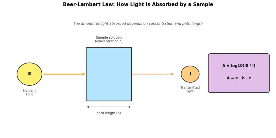
Figure 1: The Beer-Lambert Law explains how light passing through a sample gets absorbed based on concentration and path length.
UV Spectroscopy Formula
The Beer-Lambert Law is written using a simple formula:
A = e × b × c
Here is what each letter means:
- A = Absorbance (how much light was absorbed, this has no unit)
- e (epsilon) = Molar absorptivity, a constant value specific to each compound
- b = Path length of the sample cell, usually 1 cm
- c = Concentration of the sample solution
Absorbance is also linked to the intensity of light before and after it passes through the sample, using this formula:
A = log10 (I0 / I)
Where I0 is the intensity of light going into the sample, and I is the intensity of light coming out. This UV spectroscopy formula is the reason the technique can be used for both identifying a substance and measuring its concentration.
UV Spectroscopy Instrumentation
A UV spectrophotometer might look complicated from the outside, but its working parts are actually quite simple. Below is a basic UV spectroscopy diagram showing the main parts of the instrument, followed by a short explanation of each one.
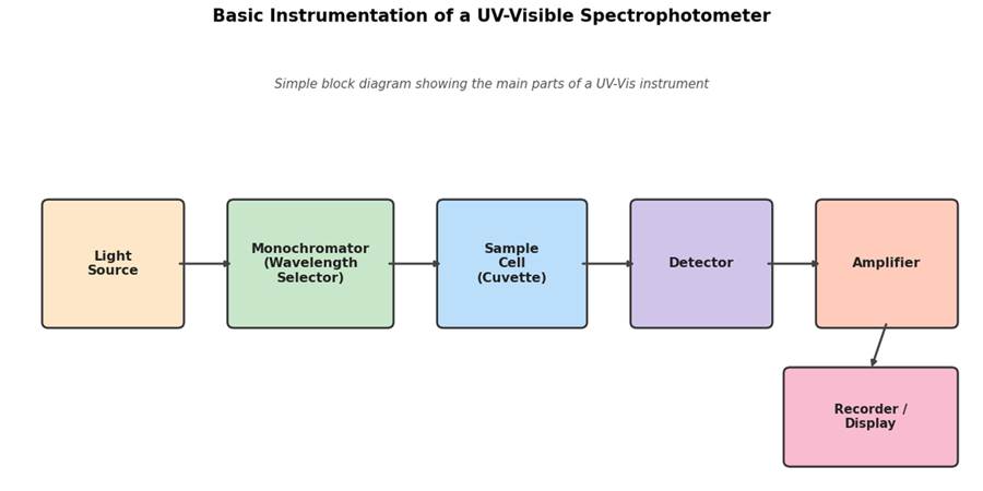
Figure 2: Basic block diagram of a UV-Visible spectrophotometer showing the light source, monochromator, sample cell, detector, and recorder.
Light Source
The light source produces the light needed for the test. Since UV spectroscopy covers both UV and visible light, two different lamps are often used. A deuterium lamp gives out UV light, while a tungsten lamp gives out visible light. Some modern instruments combine both into a single unit so the operator does not need to switch lamps manually.
Monochromator
The monochromator’s job is to pick out just one wavelength of light from the full range produced by the lamp. It usually contains a prism or a grating that splits the light into its different wavelengths, along with a slit that lets only the chosen wavelength pass through to the sample.
Sample Cell (Cuvette)
The sample is held in a small container called a cuvette. Cuvettes for UV work are made of quartz, because ordinary glass blocks UV light and would give false readings. The sample cell is usually 1 cm wide, which matches the path length used in the Beer-Lambert Law formula.
Detector
The detector measures how much light comes out the other side of the sample. It turns the light signal into an electrical signal, which is then processed by the instrument. Common detectors include photomultiplier tubes and photodiodes.
Recorder / Display
Finally, the recorder or display shows the result, either as a single absorbance value or as a full graph plotting absorbance against wavelength. Most modern instruments are connected to a computer, which stores the readings and can print out a full UV absorption spectrum.
Single Beam vs Double Beam Instruments
UV spectrophotometers generally come in two designs, and it helps to know the difference between them.
- Single beam instruments: Here, only one beam of light travels through the instrument. The blank is measured first, then the sample is measured separately in the same path. These instruments are simple and cheaper, but a little slower since you have to switch between blank and sample.
- Double beam instruments: These split the light into two beams, one goes through the sample and the other through the blank, at the same time. This design gives faster and more stable results, since it automatically corrects for small changes in lamp brightness during the test.
Most modern labs, especially in pharmaceutical analysis, prefer double beam instruments because of their better accuracy and convenience for routine testing.
UV Spectroscopy Working Principle
Now that we know the parts of the instrument, let’s look at the UV spectroscopy working principle step by step, from switching on the lamp to getting the final reading.
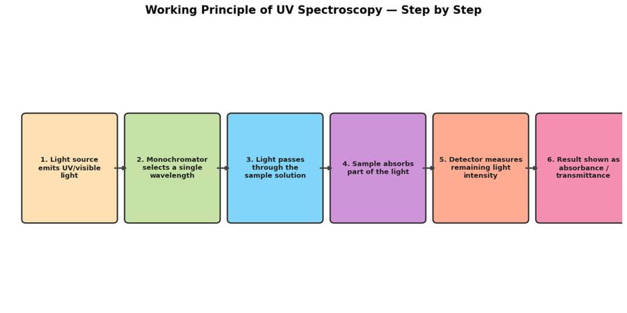
Figure 3: Step-by-step working of a UV spectrophotometer, from light generation to final result.
- The light source produces a broad beam of UV and visible light.
- This light passes through the monochromator, which selects a single wavelength.
- The selected light beam passes through the sample solution held in the cuvette.
- Part of the light is absorbed by the molecules in the sample, and the rest passes through.
- The detector picks up the light that comes out and measures its intensity.
- The instrument compares this with the original light intensity and calculates the absorbance.
- The result is shown as a number or plotted on a graph, giving the final UV absorption spectrum.
Before testing the actual sample, a blank solution (usually the solvent alone, with no sample dissolved in it) is tested first. This sets a baseline so that the solvent itself does not add any extra absorbance to the final reading.
UV Absorption Spectrum and Interpretation
A UV absorption spectrum is simply a graph. Wavelength is plotted on the horizontal axis and absorbance is plotted on the vertical axis. The graph usually shows one or more peaks, and the wavelength at the highest point of a peak is called lambda max, written as λmax.
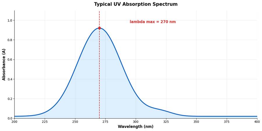
Figure 4: A typical UV absorption spectrum showing absorbance plotted against wavelength, with the peak marked as lambda max.
Chromophore and Auxochrome
A chromophore is the part of a molecule that actually absorbs UV or visible light. Common chromophores include double bonds like C=C and C=O, and ring systems like benzene.
An auxochrome is a different kind of group. It does not absorb light on its own, but when it is attached next to a chromophore, it changes how much light is absorbed and can shift the position of the peak. Groups like -OH, -NH2, and -Cl are common auxochromes.
When an auxochrome shifts the peak towards a longer wavelength, this is called a bathochromic shift, or red shift. When the peak moves towards a shorter wavelength, it is called a hypsochromic shift, or blue shift.
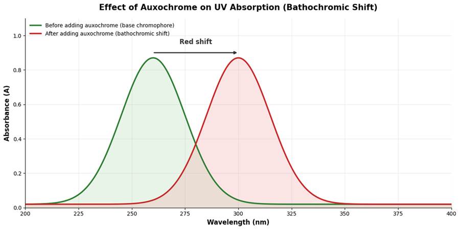
Figure 5: Adding an auxochrome group to a chromophore often shifts the absorption peak to a longer wavelength (bathochromic shift).
How to Read (Interpret) a UV Spectrum
UV spectroscopy interpretation mostly comes down to reading two things from the graph: the position of the peak (λmax) and its height (absorbance value).
- The position of λmax gives clues about the type of chromophore present in the molecule.
- The height of the peak, along with the Beer-Lambert Law, tells us the concentration of the compound.
- A shift in λmax compared to a known reference can show a change in the structure of the molecule, such as conjugation or the presence of an auxochrome.
For pure identification work, UV spectroscopy is usually combined with other techniques like IR or NMR spectroscopy, since UV data alone does not give a complete structural picture.
Factors Affecting UV Absorption
A few outside factors can change how a sample absorbs UV light, and it helps to know them so your readings stay accurate.
- Solvent: Different solvents can shift the peak position slightly. Always use a solvent that is transparent in the UV range you are working in.
- pH: Some compounds absorb light differently depending on whether they are in an acidic or basic form, so pH needs to stay consistent between tests.
- Temperature: A rise in temperature can slightly change the absorbance reading for some samples, especially if it causes the molecule to change shape.
- Concentration: Very high concentrations can bend the straight-line relationship described by the Beer-Lambert Law, so samples are often diluted before testing.
- Presence of impurities: Dust, fingerprints on the cuvette, or dissolved particles can scatter light and give a false high reading.
Keeping these factors under control is a big part of getting reliable and repeatable results from any UV spectroscopy test.
UV Spectroscopy Calibration
Like any lab instrument, a UV spectrophotometer needs regular calibration to make sure the readings it gives are accurate. UV spectroscopy calibration usually checks three main things:
- Wavelength accuracy – making sure the instrument is actually measuring at the wavelength it says it is, often checked using a holmium oxide or a standard solution with known peaks.
- Absorbance accuracy – checked using standard solutions with known absorbance values, such as potassium dichromate solution.
- Stray light – checking that no unwanted light is reaching the detector, which is usually tested using solutions like sodium nitrite or potassium chloride that block light completely at certain wavelengths.
Most labs calibrate their UV spectrophotometer at fixed intervals, such as every day before use or once a week, depending on how often the instrument is used and the standard procedures followed in that lab.
UV Spectroscopy in Pharmaceutical Analysis
One of the biggest users of UV spectroscopy is the pharmaceutical industry. UV spectroscopy in pharmaceutical analysis is used at almost every stage of drug development and quality control, from checking raw materials to testing the final tablet or injection.
- Assay of drugs: Measuring the exact amount of active ingredient present in a tablet, capsule, or injection.
- Dissolution testing: Checking how fast a tablet releases its drug into a solution, an important test for how the drug will work in the body.
- Purity checks: Comparing the UV spectrum of a sample against a known reference to spot impurities.
- Stability studies: Watching how a drug’s UV absorbance changes over time, which can show if the drug is breaking down.
- Quality control: Routine batch-to-batch testing to make sure every batch of medicine meets the required standard.
Because UV spectroscopy is fast, needs a very small amount of sample, and gives repeatable results, it remains a first-choice method in most pharmaceutical quality control labs, alongside other techniques such as HPLC.
Regulatory bodies and official pharmacopoeias, including the United States Pharmacopeia, list UV spectroscopy as an approved method for many drug assays. This is one reason pharmaceutical companies invest in well-calibrated UV instruments and trained staff, since even a small error in absorbance reading can affect whether a batch of medicine passes or fails quality control.
In a typical pharma lab, a UV method is first validated before it is used routinely. Validation checks things like accuracy, precision, linearity (how well the absorbance follows the Beer-Lambert Law across a range of concentrations), and specificity (making sure other ingredients in the formulation do not interfere with the reading). Once validated, the method can be used with confidence for daily batch testing.
UV Spectroscopy Applications
Apart from pharmaceuticals, UV spectroscopy applications spread across many fields of science and industry. Here is a quick look at where else this technique is used.
| Field | How UV Spectroscopy is Used |
| Chemistry | Identifying unknown compounds and studying reaction progress |
| Biochemistry | Measuring DNA, RNA, and protein concentration in samples |
| Food industry | Checking food color additives and detecting contamination |
| Environmental science | Testing water quality and measuring pollutant levels |
| Clinical labs | Blood and urine tests, such as measuring bilirubin or glucose |
| Cosmetics | Testing sunscreen products for how well they absorb UV rays |
| Forensic science | Analyzing ink, fibers, and other trace evidence |
UV Spectroscopy Advantages
UV spectroscopy has stayed popular for decades, and that is mostly because of these UV spectroscopy advantages:
- Simple and easy to use, even for beginners in a lab
- Fast results, often within a minute or two per sample
- Needs only a very small amount of sample
- Non-destructive in most cases, meaning the sample can still be used later
- Cost-effective compared to many other analytical techniques
- Works well for both qualitative (what is it) and quantitative (how much is it) analysis
- Widely accepted and included in official pharmacopoeia methods
UV Spectroscopy Limitations
No technique is perfect, and UV spectroscopy limitations are worth knowing before relying on it fully:
- Only works for compounds that actually absorb UV or visible light
- Cannot give full structural details of a molecule on its own
- Sensitive to impurities, dust, or fingerprints on the cuvette, which can affect readings
- Solvent choice matters a lot, since some solvents absorb UV light themselves and interfere with results
- Overlapping peaks can make it hard to analyze mixtures with several similar compounds
- Temperature and pH changes can shift the absorbance reading
Tips for Getting Accurate UV Spectroscopy Results
A few small habits in the lab can make a big difference to how reliable your UV spectroscopy results turn out to be.
- Always clean the cuvette with a lint-free tissue before placing it in the instrument, since fingerprints and dust can scatter light.
- Handle the cuvette by its frosted sides, not the clear optical faces, to avoid smudges.
- Let the lamp warm up for a few minutes before taking readings, as UV lamps can be unstable right after switching on.
- Use fresh solvent for the blank each time, and make sure it matches the solvent used for the sample.
- Check that the sample concentration falls within the linear range of the Beer-Lambert Law, diluting if needed.
- Record readings at the same wavelength (λmax) every time for consistency across a batch of tests.
These steps are simple but skipping them is one of the most common reasons students and even experienced lab workers get results that don’t match up between repeat tests.
UV Spectroscopy Experiment
Here is a simple outline of a basic UV spectroscopy experiment that is commonly done in student labs to find the concentration of an unknown sample.
- Prepare a set of standard solutions of the compound at known concentrations.
- Prepare a blank solution using only the solvent.
- Set the wavelength on the instrument to the λmax of the compound.
- Measure the absorbance of the blank first to set the baseline to zero.
- Measure the absorbance of each standard solution.
- Plot a calibration graph of absorbance versus concentration; it should form a straight line, as per the Beer-Lambert Law.
- Measure the absorbance of the unknown sample.
- Use the calibration graph to find the concentration of the unknown sample.
This basic setup is the foundation of most quantitative work done using UV-Visible spectroscopy, whether in a college lab or an industrial quality control department.
UV Spectroscopy Notes: Quick Summary
If you are revising for an exam or just want a fast recap, here are the main UV spectroscopy notes from this article in one place:
- UV spectroscopy measures how much UV or visible light a sample absorbs
- It works on the Beer-Lambert Law: A = e × b × c
- Main parts of the instrument: light source, monochromator, sample cell, detector, and recorder
- Chromophores absorb light; auxochromes shift the position of the peak
- λmax is the wavelength where absorbance is highest
- Used heavily in pharmaceutical analysis, quality control, and research labs
- Simple, fast, and low cost, but limited to light-absorbing compounds
Many students search for a UV spectroscopy PDF to study offline. You are welcome to save this page as a PDF or print it out from your browser so you have these notes handy for revision or lab reference.
UV Spectroscopy MCQs (Questions and Answers)
Here are some UV spectroscopy MCQs to test how well you have understood the topic. Try to answer before checking the correct option.
1. UV spectroscopy is based on which law?
a) Newton’s Law
b) Beer-Lambert Law
c) Ohm’s Law
d) Boyle’s Law
Answer: b) Beer-Lambert Law
2. What material is used to make a cuvette for UV work?
a) Ordinary glass
b) Plastic only
c) Quartz
d) Wood
Answer: c) Quartz
3. What is the typical wavelength range for UV light?
a) 200–400 nm
b) 700–900 nm
c) 1–10 nm
d) 1000–2000 nm
Answer: a) 200–400 nm
4. What does λmax refer to?
a) Lowest absorbance point
b) Wavelength of maximum absorbance
c) The instrument’s power rating
d) Path length of cuvette
Answer: b) Wavelength of maximum absorbance
5. Which part of the instrument selects a single wavelength of light?
a) Detector
b) Light source
c) Monochromator
d) Recorder
Answer: c) Monochromator
6. A group that shifts the absorption peak but does not absorb light itself is called:
a) Chromophore
b) Auxochrome
c) Detector
d) Solvent
Answer: b) Auxochrome
7. A shift of the absorption peak to a longer wavelength is called:
a) Hypsochromic shift
b) Bathochromic shift
c) Hyperchromic shift
d) Hypochromic shift
Answer: b) Bathochromic shift
Frequently Asked Questions (FAQ)
What is the main difference between UV spectroscopy and UV-Visible spectroscopy?
UV spectroscopy technically refers only to the ultraviolet range, but in practice, most instruments cover both UV and visible light together, which is why the term UV-Visible spectroscopy is used more often. In everyday lab talk, both names are used to mean the same instrument.
Why is a blank solution used before testing the actual sample?
The blank solution, usually just the solvent, is tested first to make sure the solvent itself is not adding any extra absorbance. This sets the reading to zero so the final result only reflects the sample, not the solvent.
Can UV spectroscopy identify an unknown compound completely?
Not on its own. UV spectroscopy gives useful clues about the type of chromophore in a molecule, but it usually needs to be combined with other methods like IR or NMR spectroscopy for a full structural picture.
Why must the cuvette be made of quartz and not glass?
Ordinary glass absorbs UV light, which would block the very light the instrument needs to measure. Quartz lets UV light pass through freely, so it does not interfere with the reading.
How often should a UV spectrophotometer be calibrated?
This depends on how often the instrument is used and the rules followed by the lab, but many labs check wavelength and absorbance accuracy daily before use, with a fuller calibration done weekly or monthly.
Is UV spectroscopy used only in pharmaceutical labs?
No. While it is very common in pharmaceutical analysis, UV spectroscopy is also used in chemistry, biochemistry, environmental testing, food quality checks, and even forensic science.
What is the difference between absorbance and transmittance?
Absorbance tells you how much light was absorbed by the sample, while transmittance tells you how much light passed through it. They are related mathematically, and most modern instruments let you switch between the two depending on what you need.
Why do we dilute a sample before measuring its UV absorbance?
At very high concentrations, the straight-line relationship in the Beer-Lambert Law can break down, leading to inaccurate results. Diluting the sample brings the absorbance into a range where the law holds true, giving a more reliable reading.
Conclusion
UV spectroscopy is one of those techniques that looks simple on the surface but is genuinely powerful once you understand what is happening inside the instrument. It works on the basic idea that molecules absorb light, and by measuring that absorption carefully, we can find out what a sample contains and how much of it is there.
From quality checks on medicines to testing water and food, UV-Visible spectroscopy plays a quiet but important role in many industries. If you are a student, understanding the principle, instrumentation, and the Beer-Lambert Law will take you a long way, not just for exams but for real lab work too.
Keep this guide handy as your go-to UV spectroscopy notes, whether you are preparing for a university exam, setting up an experiment for the first time, or just trying to understand a lab report that mentions λmax and absorbance. Once the basic principle clicks, the rest of the topic becomes much easier to follow.
People also interested in these articles
- Pharmaceutical Stability Testing Guide: How to Keep Medicines Safe and Effective
- Cold Chain in Pharmaceutical Industry: A Complete Guide
- HPLC Explained: Principle, Instrumentation, Working, Types, Applications, Validation & Interview Questions
- Dissolution Testing in Pharmaceutical Industry
- Gas Chromatography: Principles, Working, Components, Applications, Advantages & Limitations
- Pharma QA vs QC Difference
- Gas Chromatography Detectors: Types, Working Principles, and Selection Guide